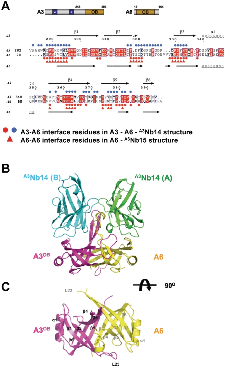
Figure 1.
Structure of heterotetramer and heterodimer. (A) Sequence alignment of A3OB and A6. A schematic representation of full-length A3 and full-length A6 with their OB-fold domain and the zinc finger domains (Z) shown in the upper panel. A3OB and A6 sequences (acronyms provided on the left) are aligned in the lower panel. Strictly conserved residues in both sequences are in red boxes. Secondary structure elements are indicated on top and below. Red and blue spheres indicate residues forming the A3OB–A6 interface; red triangles the interface residues of the A6 dimer (PDB-ID: 3K7U) (54). (B) The A3OB–A6-(A3Nb14)2 heterotetramer. The heterotetramer is shown perpendicular to the pseudo-2-fold axis relating the OB-folds of A3OB and A6. A3OB is depicted in magenta; A6 in yellow; A3Nb14 in complex with A3OB in blue; A3Nb14 in complex with A6 in green. The heterotetramer depicted is part of an [A3OB–A6–(A3Nb14)2]2 heterooctamer occurring in the crystal lattice (Supplementary Figure S7). (C) The A3OB–A6-heterodimer. A view along the pseudo-2-fold of the A3OB–A6 dimer of the heterotetramer shown in (B) is in the same color code. The N-terminal β-strands β1 of each monomer run anti-parallel to each other in the center of an extended β-sheet.

