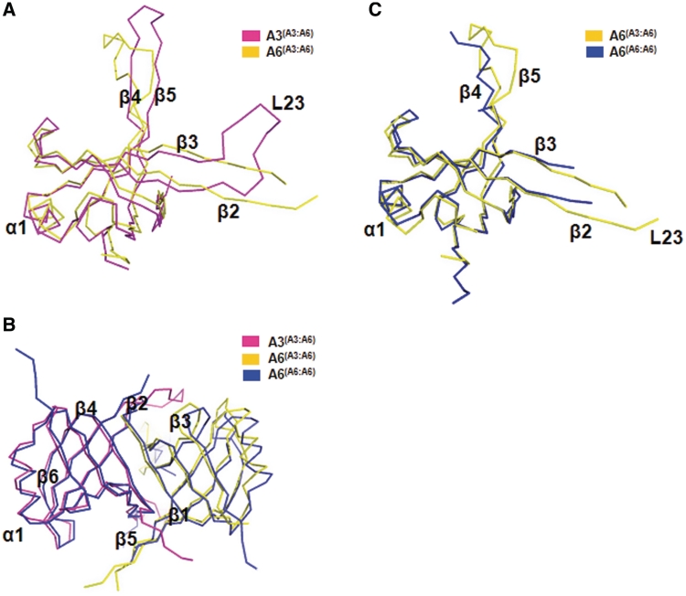
Figure 2.
Comparison of A3OB and A6. (A) Superposition of the A3OB and A6 chains from the A3OB-A6 heterodimer. The A3OB chain is shown in magenta; A6 from the A3OB–A6 heterodimer in yellow. Note the large conformational differences in the L23 loop, as well as in the β4–β5 hairpin. (B) Superposition of the A3OB–A6 and A6–A6 dimers. A view along the pseudodyad of the heterodimer. The A3OB chain is shown in magenta; A6 from the A3OB–A6 heterodimer in yellow; both A6 subunits from the A6–A6 homodimer in blue (PDB-ID: 3K7U) (54). (C) Superposition of A6 in the A3OB-A6 heterodimer and A6 in the A6 homodimer. A6 from the heterodimer is depicted in yellow; A6 from the homodimer in blue (PDB-ID: 3K7U) (54). Note that the L23 loop, as well as the β4–β5 hairpin of A6 is better ordered in the A3OB-A6 heterodimer than in the A6 dimer.

