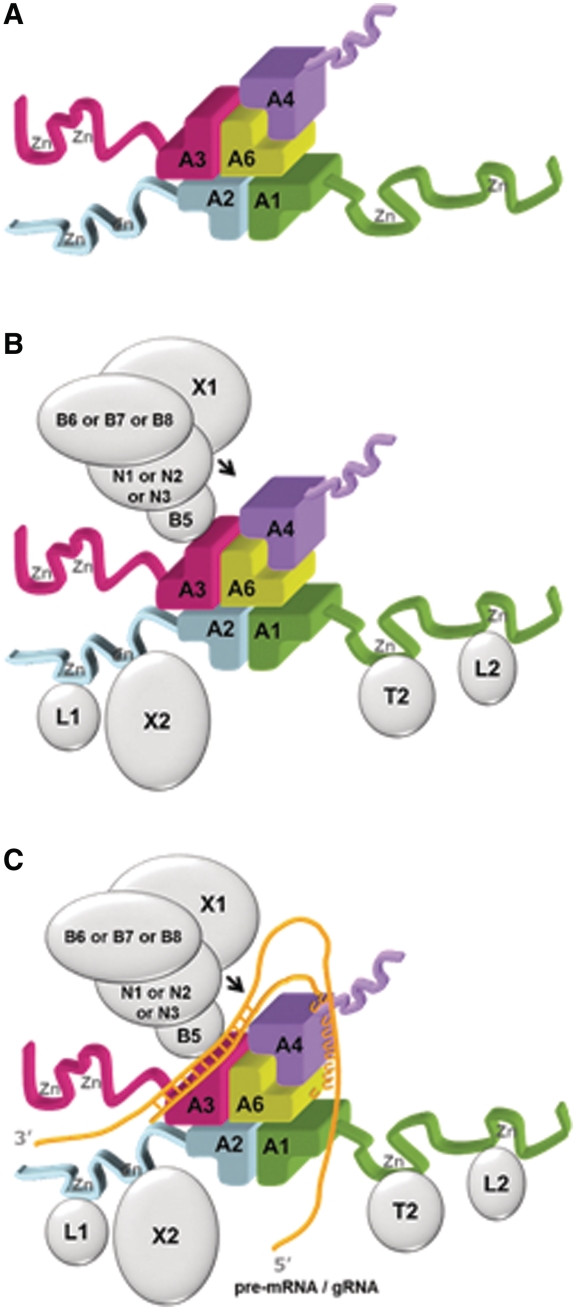
Figure 6.
An OB-fold center in the core of the editosome. (A) Model for a “five OB-fold” center of the editosome. The basis is a shifted heterotetramer (in this example, comprising the OB-folds of A1, A2, A3 and A6) plus an additional A4OB fold interacting with A6. Please note that while the A3OB–A6 heterodimer is experimentally observed, the positions of A1OB, A2OB and A4OB in this figure can be interchanged and still yield a model in agreement with the experimental constraints mentioned in the text. Therefore, the arrangement shown is one of six possible models of these five OB-folds forming the center of the editosome core. (B) The same five OB-fold center as in (A) with the additional domains in the OB-fold interaction proteins shown as ribbons and the proteins interacting with these domains outlined as silver ellipsoids. Note that three different types of editosomes have been reported to exist, containing either N1-B6, or N2-B7, or N3-B8-X1, in addition to the core (52). (C) A global outline of how a pre-mRNA•gRNA duplex might interact with the five OB-fold center shown in (A). The gold wires indicate RNA, with the duplex in the anchor region shown as crossbars, and the poly-U tail of the mature gRNA as a series of U's. The arrow points to an editing site. The surrounding enzymes might display mobility with respect to the five OB-fold center. See also text.

