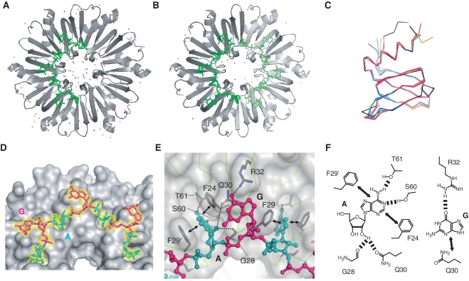
Figure 6.
Overall structures of BsHfq and the RNA aptamer, AGr. (A) Quaternary structure of BsHfq–AGr in co-crystal 1. One RNA molecule binds to BsHfq hexamer. Water molecules are shown as magenta spheres. (B) Quaternary structure of BsHfq–AGr in co-crystal 2. Symmetry-related molecule in BsHfq–AGr is shown in light gray and light green. Two RNA molecules bind to one BsHfq hexamer. Water molecules are shown as magenta and gray spheres. (C) Superposition of Cα trace of subunits from several bacterial Hfqs. Co-crystals 1 and 2 are shown in black and gray, respectively. SaHfq (PDB ID: 1KQ1) is shown in magenta, SaHfq–AU5G (PDB ID: 1KQ2) in red, EcHfq (PDB ID: 1HK9) in cyan, EcHfq–poly(A) (PDB ID: 3GIB) in blue and P. aeruginosa Hfq (PDB ID: 1U1S) in orange. Pairwise root-mean-square deviations of backbone (N, Cα, C) atoms for secondary structure elements in these subunits from co-crystal 1 structure were <0.36 Å. (D) BsHfq and AGr are shown as gray surface with cyan (A residue) or magenta (G residue) sticks, respectively. Part of 2Fo–Fc electron density map (yellow) contoured at 1.5 σ is shown for AGr. (E) Close-up view of BsHfq–AGr interface. BsHfq is shown as semi-transparent molecular surface (light gray). Polypeptide backbone is drawn as line representation (light green), while AGr is shown as ball-and-stick representations and colored as in (D). Side chains involved in RNA-binding are labeled and are shown as gray bonds colored according to atom type. Hydrogen bonding and stacking interactions between RNA and Hfq are shown as dashed lines and arrows, respectively. (F) Schematic representations of interactions between BsHfq and A/G residues in AGr. Dashed lines and arrows indicate hydrogen bonding and stacking interactions, respectively.

