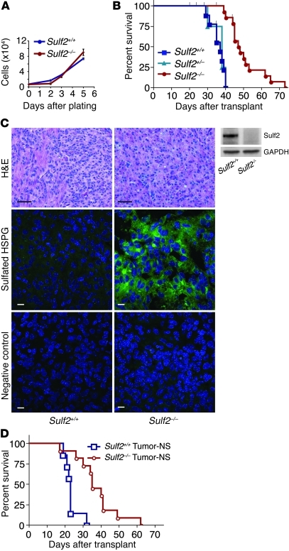
Figure 4. Prolonged survival conferred by ablation of Sulf2 in tumorigenic neurospheres.
(A) Similar in vitro growth of Sulf2+/+;Ink4a/Arf–/– or Sulf2–/–;Ink4a/Arf–/– tumorigenic neurospheres when cultured under nonadherent conditions (n = 3; mean ± SEM). (B) Kaplan-Meier survival analysis. Mice transplanted with Sulf2–/– cells have prolonged survival (median survival of 48 days) relative to mice transplanted with Sulf2+/+ or Sulf2+/– cells (median survival 37 days and 38 days, respectively, P < 0.001 for Sulf2–/– [n = 14] versus Sulf2+/+ [n = 9] or Sulf2+/– [n = 4]). 3 independent Sulf2–/– tumor progenitor lines were analyzed. Censored animals (black ticks) indicate individual mice sacrificed for tumor analysis prior to signs of tumor. (C) Similar tumor histology in Sulf2+/+ and Sulf2–/– tumors (H&E) despite absence of Sulf2 protein in Sulf2–/– tumor-NS (right panels, Western blot). Sulf2–/– tumors exhibit increased sulfated HSPGs (RB4CD12) compared with Sulf2+/+ tumors. Negative control antibody (MPB49) does not bind HSPG. Scale bars: 40 μm (H&E); 10 μm (HSPG, control). (D) Kaplan-Meier survival analysis demonstrating mice transplanted with tumor-NS isolated from Sulf2–/– tumors (median survival, 35 days) retain prolonged survival relative to those with tumor-NS from Sulf2+/+ tumors (median survival, 23 days). P < 0.001 for Sulf2–/– tumor-NS (n = 11) versus Sulf2+/+ tumor-NS (n = 7). See also Supplemental Figure 4.

