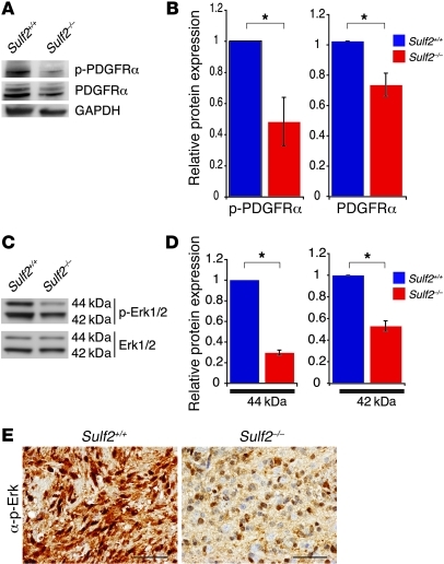
Figure 8. Sulf2 alters the activity of PDGFRα in murine tumor-NS.
(A) Phosphorylated and total PDGFRα levels in Sulf2+/+ and Sulf2–/– tumor-NS. Western blots were probed for GAPDH as a loading control. (B) Quantification of p-PDGFRα and total PDGFRα levels in tumor-NS from Sulf2+/+ and Sulf2–/– cells normalized to mean ± SEM (n = 3 independent experiments). *P < 0.05. (C) SULF2 also affected the activity of downstream signaling pathways. Phosphorylated and total Erk1/2 (p44/p42) levels in Sulf2+/+ and Sulf2–/– tumor-NS. (D) The relative mean ratio of phosphorylated Erk to total Erk levels in Sulf2+/+ and Sulf2–/– tumor-NS normalized to Sulf2+/+ levels ± SEM (n = 3 independent experiments). *P < 0.005. (E) Sulf2+/+ tumors had more prominent phosphorylated Erk immunostaining relative to Sulf2–/– tumors. Scale bars: 50 μm.

