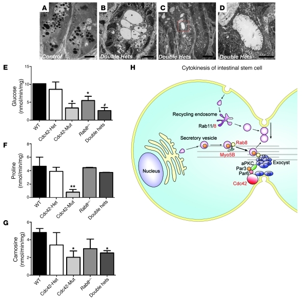
Figure 8. Cdc42 and Rab8a double heterozygous mice show abnormal crypt morphology and epithelial uptake.
(A–D) TEM micrographs of control and double heterozygous crypts. Scale bars: 10 μm (A–C); 500 nm (D). (E–G) Intestinal transporter uptake assays for glucose, proline, and carnosine. *P < 0.05 compared with wild type; #P < 0.05 compared with wild type and single heterozygous; **P < 0.01 compared with wild type. (H) A model depicting the coordination of Cdc42 and Rab8a during intestinal stem cell division and epithelial morphogenesis. Cdc42, Rab8a, and Myo5B are shown in red to indicate their association with MVID. The exocyst, an octameric protein complex of Sec3, Sec5, Sec6, Sec8, Sec10, Sec15, Exo70, and Exo84, is indicated as blue circles at midbody.

