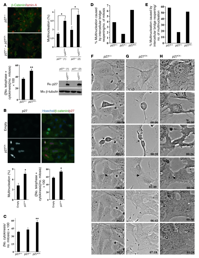
Figure 3. p27CK– causes a cytokinesis defect.
(A) Multinucleation was quantified (500 cells counted/line, n = 3) in two lines of immortalized p27–/– MEFs infected or not with p27CK– and stained for β-catenin and lamin-A (original magnification, ×400). p27CK– levels following retroviral infection were measured by immunoblot for p27, and β-tubulin levels were used as loading control. Lower graph: For each cell line, the proportion of mitotic cells in telophase and cytokinesis was determined in 50–100 mitotic figures; results are mean of 5 independent experiments. (B) Overexpression of p27CK– in HeLa cells causes multinucleation. HeLa cells were transfected with either empty vector or a vector encoding p27CK–. Transfected cells were visualized by p27 staining and co-labeled for β-catenin, and 600 p27-positive cells were counted (original magnification, ×400). Graphs show the average number of multinucleated cells in 3 independent experiments. Right graph: number of mitotic cells in telophase and cytokinesis observed in 100 mitotic figures (n = 4). In A and B, *P < 0.05, **P < 0.01, Student’s t test. (C) Number of mitotic cells in cytokinesis in immortalized p27+/+, p27–/–, and p27CK– MEFs. One hundred mitotic figures were counted per genotype in 3 independent experiments. Results were analyzed by 1-way ANOVA with the Tukey-Kramer multiple comparison test; p27+/+ versus p27CK–, **P < 0.01. Error bars in A–C represent SEM. (D–H) Videomicroscopy of p27+/+, p27–/–, and p27CK– MEFs undergoing mitosis. (D) Incidence of multinucleation caused by cytokinetic regression relative to the total number of mitotic figures recorded (p27+/+: n = 181; p27–/–: n = 117; p27CK–: n = 243). (E) Nature of the defect causing multinucleation: multinucleation caused by reopening of the intercellular bridge as a percentage of the total number of multinucleation was determined for each genotype. (F–H) Examples of phase contrast videomicroscopy of mitotic p27+/+ (F) and p27CK– MEFs (G and H). Arrows indicate the intercellular bridge. In p27CK–, cells undergo mitosis normally until the formation of intercellular bridge, which regresses to form binucleated cells. Original magnification, ×200.

