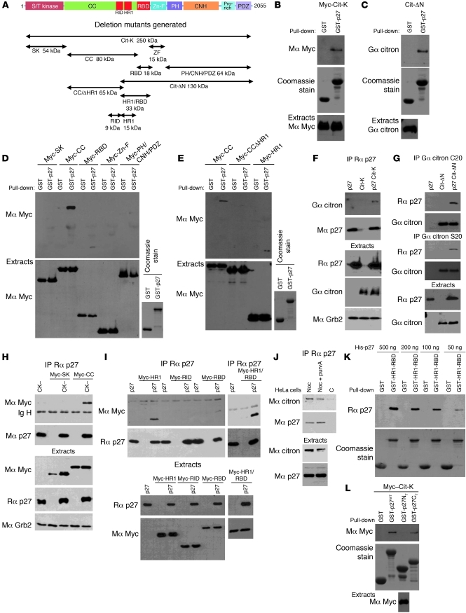
Figure 4. The C-terminal half of p27 interacts with the HR1 domain of citron-K.
(A) Schematic representation of citron-K (Cit-K) and deletion mutants. All constructs except the Cit-ΔN mutant contain an N-terminal Myc epitope tag. S/T kinase, Ser/Thr kinase domain; CC, CC domain; Zn-F, Zn finger; PH, pleckstrin homology; CNH, citron homology; Pro-rich, proline-rich region: PDZ, PDZ binding domain. (B–E) HEK293 cells transfected with citron-K or the indicated deletion mutant were subjected to pull-down assays using GST or GST-p27 beads. The amounts of citron-K or deletion mutant bound to the beads and of transfected protein present in the extracts were detected by immunoblot using the indicated antibodies (Mα, mouse anti-; Gα, goat anti-; Rα, rabbit anti-). The amount of GST or GST-p27 used in the assays was visualized by Coomassie staining. (B and C) p27 interacts with citron-K. (D) p27 interacts with the CC domain of citron-K and more weakly with the RBD domain. (E) The HR1 domain present in the CC domain is necessary for p27 interaction. (F–I) Co-immunoprecipitations using rabbit anti-p27 (C19) (F, H, and I) or two different goat anti-citron antibodies (C20 or S20) (G). Co-immunoprecipitated proteins were detected by immunoblot with the indicated antibodies. The immunoprecipitated proteins were visualized by reprobing for mouse anti-p27 (F8) (F, H, and I) or goat anti-citron (C20) (G). Immunoblots of extracts show the level of transfected proteins in each condition. Grb2 levels were used as loading control. (F and G) p27 interacts with citron-K in vivo. (H and I) p27 interacts with all mutants containing the HR1 domain. A weaker interaction is also seen with the RBD. (J) p27 interacts with citron-K in HeLa cells. Endogenous p27 was immunoprecipitated using rabbit anti-p27 (C19) in HeLa cells. The co-immunoprecipitated citron-K was visualized using mouse anti–citron-K antibody. Endogenous levels of each protein are shown in the lower panels. Noc, nocodazole; purvA, purvalanol A; C, control. (K) p27 interacts with citron-K directly. Different amounts of recombinant His-p27 were used in pull-down assays with GST or GST-HR1-RBD beads. Immunoblot show the amount of p27 bound to the beads. The amount of GST or GST-HR1-RBD used in the assay was visualized by Coomassie staining. (L) The C-terminal half of p27 interacts with citron-K. Pull-downs were realized as in B with GST-p27WT, GST-p27NT (N-terminal, aa 1–86), and GST-p27CT (C terminal, aa 87–198).

