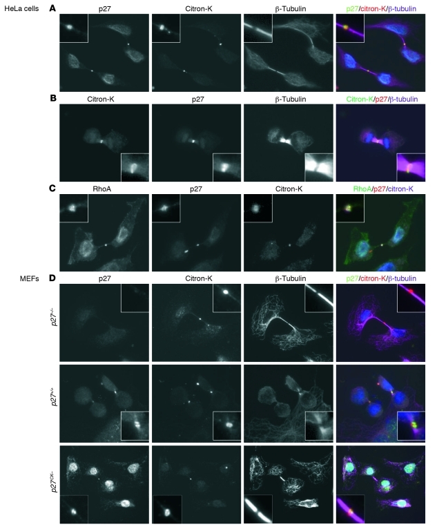
Figure 5. Colocalization of p27 and citron-K at the mid-body.
(A and B) After 24 hours in culture, HeLa cells were permeabilized with digitonin (40 μg/ml) before fixation. Mid-bodies were visualized with citron-K (goat anti–citron C20) staining in red (A) or green (B); p27 was detected with rabbit anti-p27 (C19) antibody in green (A) or red (B); and microtubules were stained with mouse anti–β-tubulin (purple) (original magnification, ×600). (C) HeLa cells were permeabilized with digitonin prior to fixation and stained with mouse anti-RhoA (26C4) antibody (green), rabbit anti-p27 (C19) (red), and goat anti-citron (C20) (purple) (original magnification, ×600). (D) Primary MEFs derived from p27+/+, p27–/–, and p27CK– mice were stained with anti-p27 (C19), anti-citron (C20), and anti–β-tubulin after digitonin permeabilization and fixation (original magnification, ×600).

