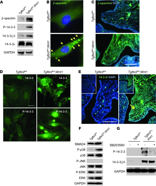
Figure 2. Identification of molecules with upregulated expression in primary MEPM cells from Tgfbr2fl/fl;Wnt1-Cre mice.
(A) Immunoblotting analysis of indicated molecules in primary MEPM cells of Tgfbr2fl/fl and Tgfbr2fl/fl;Wnt1-Cre mice. (B) Immunofluorescence analysis of primary MEPM cells of Tgfbr2fl/fl and Tgfbr2fl/fl;Wnt1-Cre mice using anti–β-spectrin antibody. Arrows indicate expression of β-spectrin. Original magnification, ×400. (C) Immunohistochemical staining of β-spectrin and DAPI staining in sections of Tgfbr2fl/fl and Tgfbr2fl/fl;Wnt1-Cre mice at E14.0. Scale bar: 50 μm. (D) Immunofluorescence analysis of primary MEPM cells from Tgfbr2fl/fl and Tgfbr2fl/fl;Wnt1-Cre mice using anti–14-3-3ζ/δ (14-3-3) or anti–phosphorylated 14-3-3 (P-14-3-3) antibody. Scale bar: 20 μm. (E) Immunohistochemical staining of 14-3-3ζ/δ and DAPI staining in sections of Tgfbr2fl/fl and Tgfbr2fl/fl;Wnt1-Cre mice at E13.5. 14-3-3ζ/δ expression appears increased in Tgfbr2fl/fl;Wnt1-Cre palate (arrow) compared with that in Tgfbr2fl/fl littermate control. Scale bar: 50 μm. Insets show lower-magnification images (original magnification, ×100). (F) Immunoblotting analysis of indicated molecules in primary MEPM cells from Tgfbr2fl/fl and Tgfbr2fl/fl;Wnt1-Cre mice. P-p38, phosphorylated p38; P-JNK, phosphorylated JNK; P-ERK, phosphorylated ERK. (G) Immunoblotting analyses of indicated molecules in primary MEPM cells of Tgfbr2fl/fl and Tgfbr2fl/fl;Wnt1-Cre mice treated with (+) or without (–) p38 MAPK inhibitor SB203580. P-14-3-3, phosphorylated 14-3-3.

