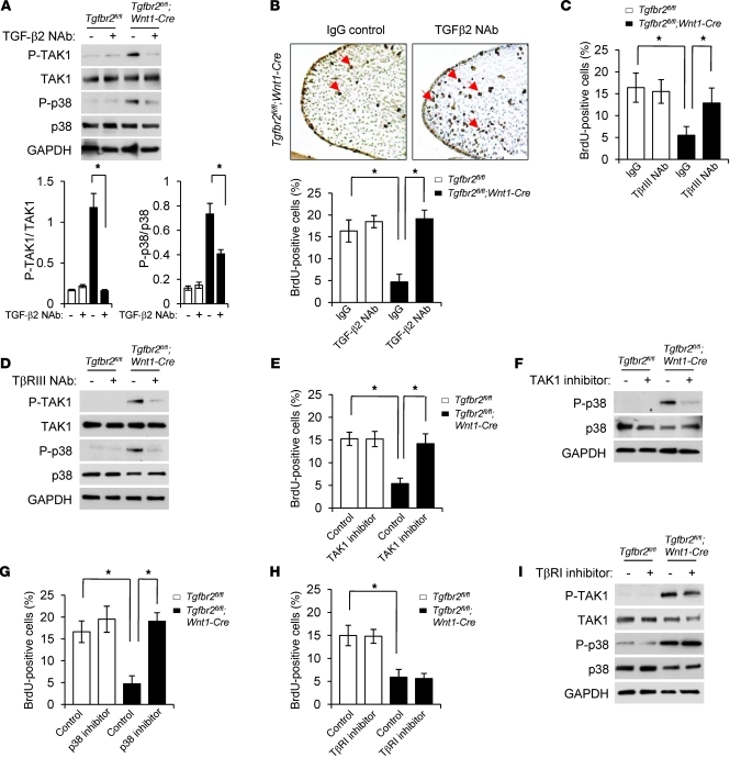
Figure 5. Inhibition of TGF-β2/TβRI/TβRIII–mediated TAK1/p38 MAPK activation rescues a cell proliferation defect in Tgfbr2fl/fl;Wnt1-Cre palates.
(A) Immunoblotting analysis of indicated molecules in MEPM cells from Tgfbr2fl/fl and Tgfbr2fl/fl;Wnt1-Cre mice cultured with (+) or without (–) TGF-β2 NAb. The bar graphs show the ratios of indicated molecules after quantitative densitometry analysis of immunoblotting data. *P < 0.05. (B) BrdU incorporation in the palate of Tgfbr2fl/fl;Wnt1-Cre mice after IgG or TGF-β2 NAb treatment. Arrows indicate BrdU-positive cells. Original magnification, ×200. The bar graph shows the percentage of BrdU-labeled nuclei in the indicated genotyping palate treated with TGF-β2 NAb or IgG. *P < 0.05. (C) Quantitation of the percentage of BrdU-labeled nuclei in the indicated genotyping palates treated with TβRIII NAb or IgG. *P < 0.05. (D) Immunoblotting analysis of indicated molecules in indicated genotyping MEPM cells cultured with (+) or without (–) TβRIII NAb. (E) Quantitation of the percentage of BrdU-labeled nuclei in the indicated genotyping palates treated with TAK1 inhibitor or vehicle. *P < 0.05. (F) Immunoblotting analysis of indicated molecules in indicated genotyping MEPM cells cultured with (+) or without (–) TAK1 inhibitor. (G) Quantitation of the percentage of BrdU-labeled nuclei in the indicated genotyping palates treated with p38 MAPK inhibitor or vehicle. *P < 0.05. (H) Quantitation of the percentage of BrdU-labeled nuclei in the indicated genotyping palates treated with TβRI kinase inhibitor or vehicle. *P < 0.05. (I) Immunoblotting analysis of indicated molecules in primary indicated genotyping MEPM cells cultured with (+) or without (–) TβRI kinase inhibitor or vehicle.

