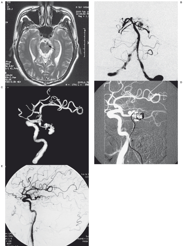
Figure 2.
Case 9. A) A T2-weighted MRI showed a flow void at the basil cistern with compression of the left peduncle. B) Left vertebral injection showed the dissecting aneurysm and aplastic P1 segment. C) Left carotid artery injection showed the aneurysm was opacified better from anterior circulation. D) Trans-vertebral embolization under the roadmap from LICA injection. E) Postprocedural angiogram showed the aneurysm were completely occluded

