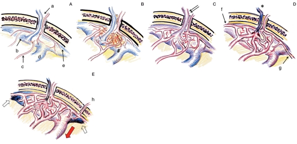
Figure 2.
Mechanism of development of the DAVF at the superior sagittal sinus (SSS) as a representative of the sinus type DAVF. A) Normal site. Emissary vein (EV) and artery (a) is penetrating through a foramen of the parasagittal skull. EV is connected with the venous lacunae (b). Meningeal arteries (c) have no connection with SSS (d) and cortical vein (e). B) Neovascularization (arrow) and vessel dilatation induced by dural inflammation (#). C) AV shunt formation at the level of dural arteriole and penetration into the sinus (initial stage of DAVF). Note the shunt flow draining into the sinus as well as EV (double arrow). D) Shunt development with thrombosis of an emissary vein (asterisk) and recruitment of distal arteries from anterior falx artery (f) and posterior meningeal arteries (g). E) Maturation of DAVF with the reflux to cortical veins (red arrow) due to sinus occlusion (white arrow). Note the further recruitment of feeders from the other side or transosseous branches (h).

