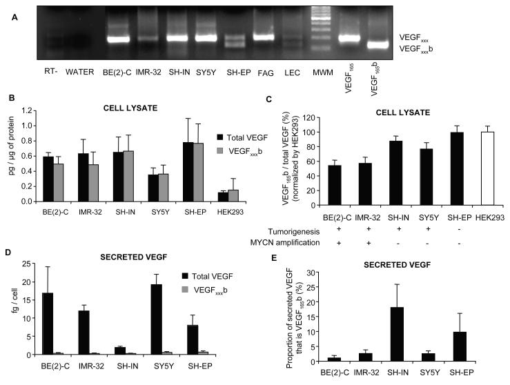
Figure 3. Differential expression and secretion of VEGF isoforms by neuroblastoma cell lines.
(A) Extraction of mRNA from NB cell lines followed by RT-PCR using primers to detect both families of isoforms. FAG is fetal adrenal gland and LEC, lymphatic endothelial cells. (B) ELISA for VEGFxxxb and total VEGF on cell lysates for different NB cell lines where MYCN oncogene amplification is associated with poor prognosis. Results normalized to protein. HEK293 cells were used as positive control for the expression of VEGF xxxb. (C) VEGFxxxb relative expression decreases significantly in highly tumorigenic, MYCN amplified cell lines (One-way ANOVA, post test for linear trend, p<0,01). Results normalized to HEK293 cells. (D) Cell ELISA for VEGFxxxb and total VEGF showing a significant secretion of VEGF (1-20fg/cell) in all neuroblastoma cell lines. VEGFxxxb secretion was 1-2 orders of magnitude lower (0.2-0.6fg/cell). Results normalized by cell number. (E) Proportion of secreted VEGF that is VEGFxxxb. Only SH-IN and SH-EP cells secreted more than 5% of its VEGF as VEGFxxxb.

