
Fig. 1.
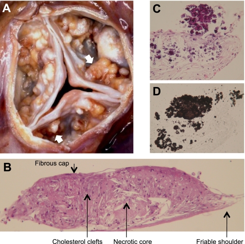
Calcific atherosclerotic nodules. A: gross pathology of human calcific aortic valve stenosis, showing numerous calcific nodules (arrows) on the aortic face of the valve leaflets (kindly provided by Michael Fishbein, UCLA School of Medicine). B: histochemical staining of calcifying vascular cell (CVC) nodule produced in vivo within a subcutaneous diffusion chamber implanted in a hyperlipidemic ApoE-deficient mouse. C and D: high-magnification images of serial sections from the shoulder region revealing calcium mineral deposits by hematoxylin and eosin (C) and von Kossa staining (D). Nodules measure approximately 0.5–2 mm in diameter.