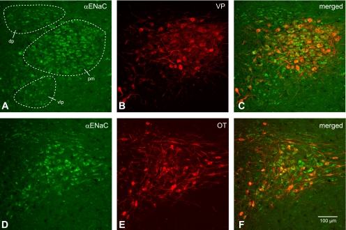
Fig. 3.

Colocalization of α-ENaC subunit and VP-NP immunoreactivity in coronal section of the rat paraventricular nucleus (PVN). A and D: α-ENaC subunit immunoreactivity labeled with DyLight 488-conjugated secondary antibody. Prominent α-ENaC immunoreactivity is observed in somata and proximal parts of dendrites of MNCs within the PVN. Most of these α-ENaC-immunoreactive magnocellular cells are located in a cluster of cells in the posterior magnocellular (pm) region. The parvocellular cells in dorsal (dp), ventrolateral (vlp), and posterior parvocellular regions are also immunoreactive to α-ENaC subunit. B: VP-NP immunoreactivity labeled with DyLight 594-conjugated secondary antibody in same section and image plane as in A. Note that the VP-NP immunoreactive cells form a cluster in the lateral portion of the PVN. E: OT-NP immunoreactivity labeled with DyLight 594-conjugated secondary antibody in same section and image plane as in D. C and F: the merged images revealed that α-ENaC immunoreactivity is colocalized with both VP and OT immunoreactivities within MNCs in the PVN. Note that the lateral cluster of the cells is composed mostly of VP. MNCs are immunoreactive to α-ENaC as well as those sparsely located OT MNCs around these VP neurons. However, the parvocellular cells immunoreactive to α-ENaC within dp, vlp, and posterior parvocellular regions are not immunoreactive to VP or OT.
