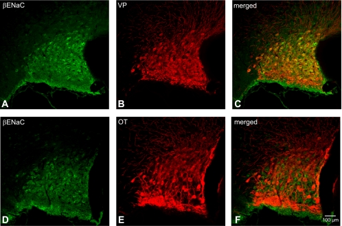
Fig. 4.

Colocalization of β-ENaC subunit with both VP-NP and OT-NP immunoreactivity in coronal section of the rat SON. A and D: β-ENaC subunit immunoreactivity labeled with DyLight 488-conjugated secondary antibody. Note that immunoreactivity of β-ENaC in the SON is confined within the somata of MNCs. B: VP-NP immunoreactivity labeled with DyLight 594-conjugated secondary antibody in same section and image plane as in A. E: OT-NP immunoreactivity labeled with DyLight 594-conjugated secondary antibody in same section and image plane as in D. C and F: the merged images reveal that these β-ENaC-immunoreactive neurons are all VP and not OT immunoreactive neurons.
