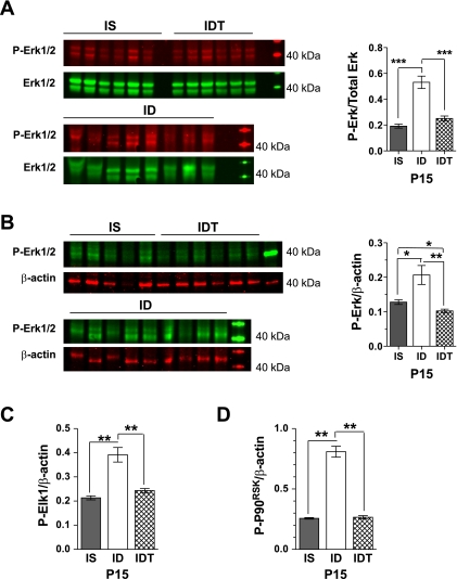
Fig. 4.
Increased phosphoinositide 3-kinase (PI3K)-independent extracellular signal-regulated kinase (Erk) 1/2 signaling in P15 ID hippocampus. A and B: Western blot images showing P-Erk1/2 with respective total Erk1/2 (A) and P-Erk1/2 with respective β-actin (B). Corresponding quantified data are shown on right. C and D: increased phosphorylated levels of Elk1 (C) and P-p90rsk (D), downstream effectors of P-Erk1/2 signaling. Values are means ± SE; n = 4–9/group. *P < 0.05, **P < 0.01, and ***P < 0.001.

