
Fig. 5.
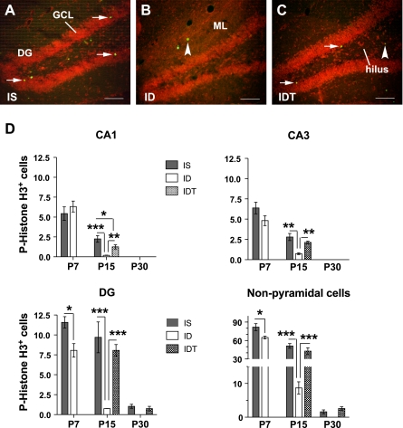
Reduced neurogenesis in ID hippocampus. A–C: representative P15 hippocampus showing phosphorylated histone H3 (pH3-ir/P-H3+, green) immunoreactive cells in dentate gyrus (DG) granular cell layer (GCL, arrows) and in noncell layer (arrowheads). Sections were counterstained with propidium iodide (red). Molecular layer (ML), scale bar = 100 μm. D: quantified P-H3+ cells found within CA1, CA3, and DG (including subgranular layer) and outside of pyramidal or granular cell layers (nonpyramidal cells). No P-H3+ cell was found in P30 hippocampal CA1 or CA3. Values are means ± SE; n = 3 (ID) and 6 (IS and IDT). *P < 0.05, **P < 0.01, and ***P < 0.001.