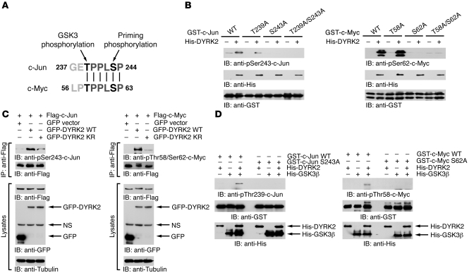
Figure 2. DYRK2 phosphorylates c-Jun and c-Myc at the priming phosphorylation residues.
(A) Sequence alignments around phosphorylation residues of c-Myc and c-Jun. (B) Recombinant DYRK2 was incubated with GST–c-Jun210–310 or GST–c-Myc1–100 in the presence of ATP. Reactants were analyzed by immunoblotting with anti–phospho–c-Jun(Ser243) (left upper panel), anti–phospho–c-Myc(Ser62) (right upper panel), anti-His (middle panels), or anti-GST (lower panels). (C) 293T cells were cotransfected with GFP vector, GFP-DYRK2 WT, or GFP-DYRK2-KR and Flag-tagged c-Jun (Flag–c-Jun) or c-Myc (Flag–c-Myc). Lysates were immunoprecipitated with anti-Flag agarose. Immunoprecipitates were then subjected to immunoblot analysis with anti–phospho–c-Jun(Ser243) (left top panel), anti–phospho–c-Myc(Thr58/Ser62) (right top panel), or anti-Flag (2nd panels). Lysates were also subjected to immunoblot analysis with anti-Flag (3rd panels), anti-GFP (4th panels), or anti-tubulin (bottom panels). (D) Recombinant DYRK2 and/or His-GSK3β were incubated with GST–c-Jun210–310 or GST–c-Myc1–100. Reaction products were analyzed with the indicated antibodies.

