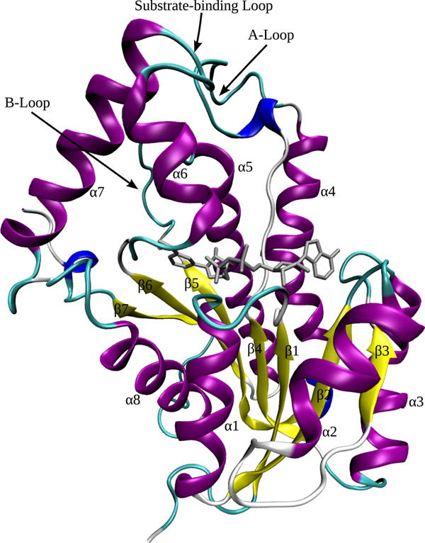
Figure 1.
The InhA tertiary structure. Ribbons representation of Mtb’s InhA (PDB ID:1ENY) rigid receptor model (colored by secondary structure) in complex with the coenzyme NADH (in metallic grey). In yellow are the 7-strands parallel β-sheet and in magenta the eight α-helices, connected by loops (in cyan) and turns (in white). The protein’s N-terminus is composed by helices α1 and α2, and by the β1 to β3 strands, while the C-terminus is formed by helices α7 and α8. Figure produced with VMD [29].

