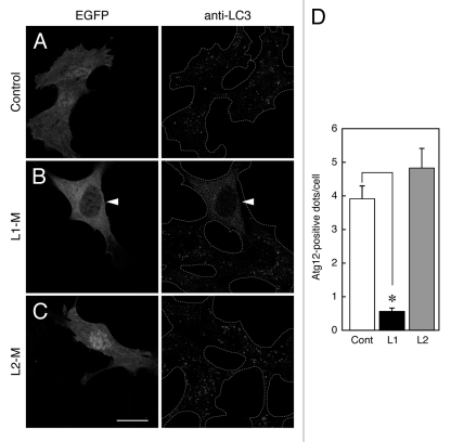
Figure 7.
Effect of overexpression of Atg16Ls-M on phagophore formation. (A–C) MEF cells transiently expressing EGFP-Atg16L1-M, EGFP-Atg16L2-M, or EGFP alone were cultured in HBSS for 1.5 h and fixed in 4% paraformaldehyde. The cells were stained with anti-LC3 antibody. The arrowheads indicate EGFP-Atg16L1-M expressing MEF cells showing inhibition of autophagosome formation. Scale bar, 20 μm. (D) Decreased number of Atg12-positive dots in cells expressing Atg16L1-M, but not Atg16L2-M, under starvation conditions. MEF cells cultured in HBSS for 1.5 h were fixed and stained with anti-Atg12 antibody. The number of Atg12-positive dots in the cells expressing Atg16Ls-M was counted. Bars represent the means and SE of one representative experiment (n > 100). *p < 0.001 (Mann-Whitney test). Similar results were obtained in an independent experiment.

