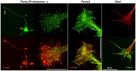
Figure 13. Hippocampal pyramidal neurons and/or their axonal growth cones labeled with antibodies to proteins involved in proteasomal degradation (top row).
The antibody specificities are indicated above. Additional filamentous-actin label is shown in the bottom row. pk, neuronal perikaryon; large arrow, axonal growth cone; small arrow, Psma-positive, large puncta.

