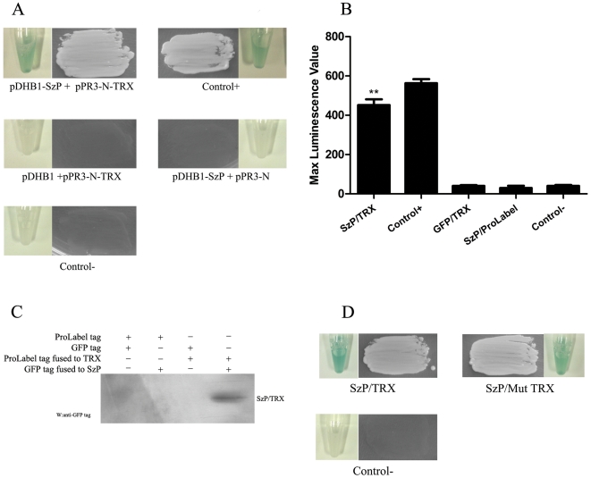
Figure 1. The interaction between SzP and TRX.
A: The split-ubiquitin yeast two hybrid assay. SzP was cloned in frame into the yeast expression vector (pDHB1), fused at its N-terminus to a small membrane anchor (the yeast ER protein Ost4) and at its C-terminus to a reporter cassette composed of the C-terminal half of ubiquitin (Cub) and a transcription factor (LexA-VP16). The recombinant TRX-pPR3-N (fusion to the mutated N-terminal half of ubiquitin) and SzP-pDHB1 were co-transformed into the NMY51 yeast strain. The co-transformed cells were selected on Trp-/Ade-/His-/Leu- 80 mM Aminotriazole dropout plates. The control+ represented co-transformation of pDSL-Delta-p53 and pDHB1-largeT (Dualsystems Biotech, Switzerland). Tubes showed the results of Liquid β-galactosidase assay. B: Luminescence value of the substrate corresponding to the pProLabel tag fused to TRX. Histogram showed max luminescence value through 1 h monitoring (n = 3, mean±SD, ** indicates that a value was significantly different (p<0.01)from the control- group). C: SzP was immunoprecipitated from HEK293 cells expressing TRX. HEK293 lysates were immunoprecipitated with rabbit polyclonal antibodies against TRX. The immunoprecipitates were subjected to the western blotting analysis with GFP monoclonal antibodies against GFP -SzP. D: pDHB1-SzP and pPR3-N-mut-TRX were co-transformed into the yeast strain NMY51. SzP interacted with mutant TRX, which had mutations Cys32 and Cys35 to Ser (C32S/C35S) in its active site. Tubes showed the results of Liquid β-galactosidase assay.

