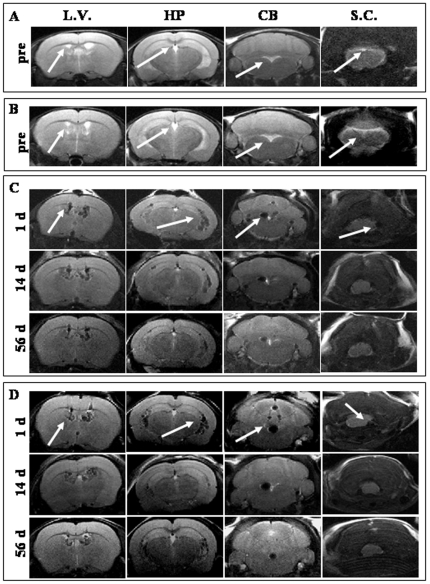
Figure 6. MRI of labeled hAFCs. Representative pictures of MRI of healthy (A) and wobbler (B) mice one day before hAFC transplantation.
Axial MRI analysis of the same healthy (C) and wobbler mice (D) at 1, 14 and 56 days after graft. For each panel, the coronal slices are indicative of different regions of the ventricular system including the site close to cell administration (L.V.), the region corresponding to the ventral hippocampus (HP), the region between the brainstem and the cerebellum (CB) and the cervical spinal cord region (S.C.).

