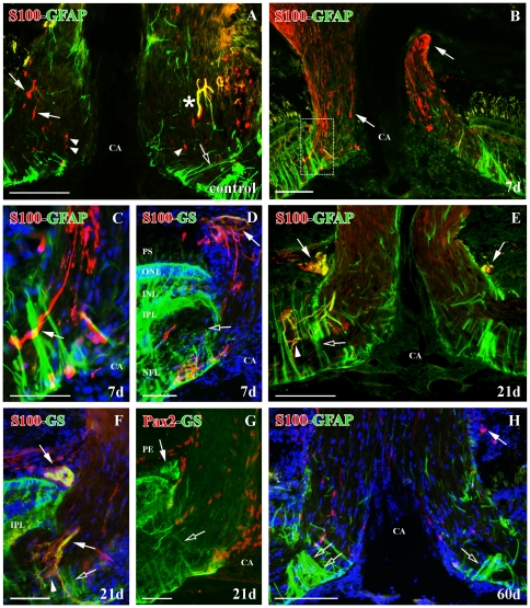
Figure 7. Double immunolabeling S100 (red)-GFAP (green), S100 (red)-GS (green) and Pax2 (red)-GS (green) with DAPI (blue) in the ONH.
(A) S100+ astrocytes (arrows), S100+/GFAP+ astrocyte (asterisk), S100+ oligodendrocytes (arrowheads) and GFAP+ Müller cell vitreal processes (empty arrow) in control goldfish. (B) S100+ astrocytes with long processes at 7 d post-injury (arrows). Square enlarged in C. (C) Detail of the S100+ astrocytes with long processes (arrow) in the optic disc (D) S100+/GS+ astrocytes surrounding the posterior part of the ONH (arrow) and GFAP+ Müller cells with disorganized vitreal processes (empty arrow) at 7 d post-injury. (E) S100+/GFAP+ astrocytes surrounding the posterior part of the ONH and in the optic disc (arrows), S100+/GFAP+ processes (arrowhead) and GFAP+ Müller cells with disorganized vitreal processes (empty arrow) at 21 d post-injury. (F) S100+/GS+ astrocytes surrounding the posterior part of the ONH and in the optic disc (arrows), S100+/GS+ processes (arrowhead) and GS+ Müller cells with disorganized vitreal processes (empty arrow) at 21 d post-injury. (G) Pax2−/GS+ astrocytes surrounding the posterior part of the ONH (arrows) and GS+ Müller cells vitreal processes (empty arrow) at 21 d post-injury. (H) Few S100+ astrocytes are surrounding the posterior part of the ONH (arrow) and there is a great amount of GFAP+ Müller cell vitreal processes (empty arrows) at 60 d post-injury. Scale bars: A–B, E, H: 100 µm; C–D, F–G: 50 µm. CA: central artery; INL: inner nuclear layer; IPL: inner plexiform layer; NFL: nerve fiber layer; ONL: outer nuclear layer; PE: pigmentary epithelium; PS: photoreceptor segments.

