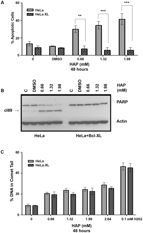
Figure 3. HAP-induced apoptosis occurs through the intrinsic pathway.
(A) Protection from HAP-induced apoptosis by overexpression of Bcl-xL. Both HeLa and HeLa-xL cell lines were treated with increasing doses of HAP for 48 hours and the percentage of apoptotic cells was determined by Hoechst staining. By two-way ANOVA, pcell line<0.0001 and pconcentration = 0.009. **p<0.01,***p<0.001 by Bonferroni multiple comparison post test for column analysis comparing means for HeLa vs. HeLa-xL hours. (B) Confirmation of protection from HAP-induced apoptosis by Bcl-xL overexpression by immunoblot for PARP cleavage. HAP induced dose-dependent cleavage of PARP in regular HeLa cells but not in HeLa cells overexpressing Bcl-xL. PAPR cleavage product is marked as cl89. (C) HAP treatment results in similar levels of DNA breaks in HeLa and HeLa+Bcl-xL cell lines (p>0.05).

