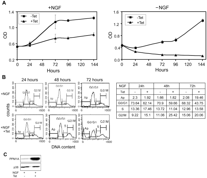
Figure 5. PPM1A overexpression and PC6-3 cells differentiation.
( A ) Cell proliferation assay. PC6-3 PPM1A wt cells were incubated with or without NGF and with or without Tet. The cells were then assayed for viability at the indicated times using XTT assay. The average of 6 different wells ± S.D. from a representative experiment was plotted. ( B ) FACS analysis of PC6-3 cells preincubated with Tet for 24 hours followed by addition of NGF for the indicated times. The table represents % of total counted cells of one experiments out of three performed. ( C ) Western blot analysis of extracts from cells treated for 144 hours with NGF with or without Tet, and assayed for PPM1A expression level with anti-PPM1A antibodies.

