
Figure 1.
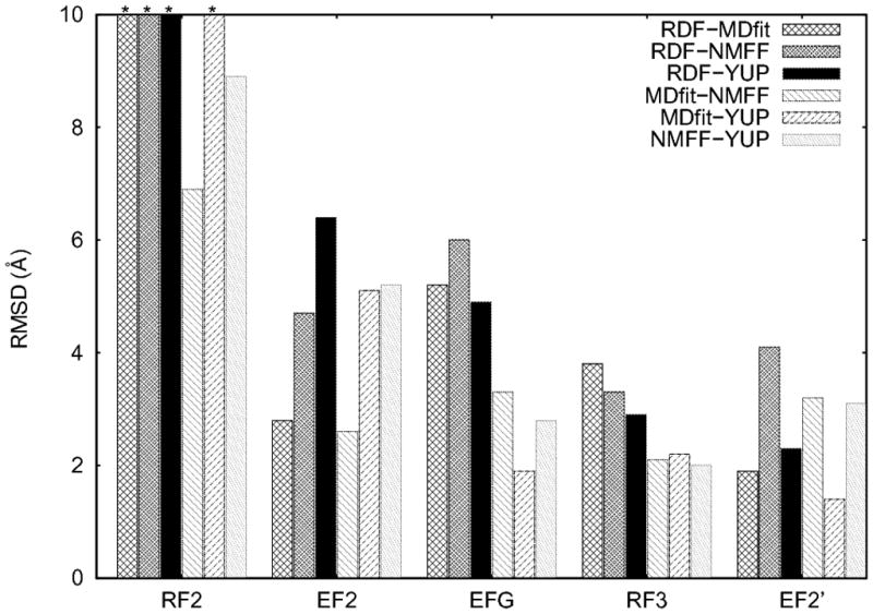
RMSD between different RDF/flexible-fitted structures of the four studied proteins obtained from different flexible-fitting approaches (MDfit, YUP.SCX, and NMFF) and from the PDB database fitted using rigid -body fitting (RDF) into Cryo-EM maps. For RF2, bars with * on the top have RMSD values > 10 Å (i.e., 14.6, 15.6, 17.8 and 11.6 Å between RDF and MDfit, RDF and NMFF, RDF and YUP, and MDfit and YUP, respectively).