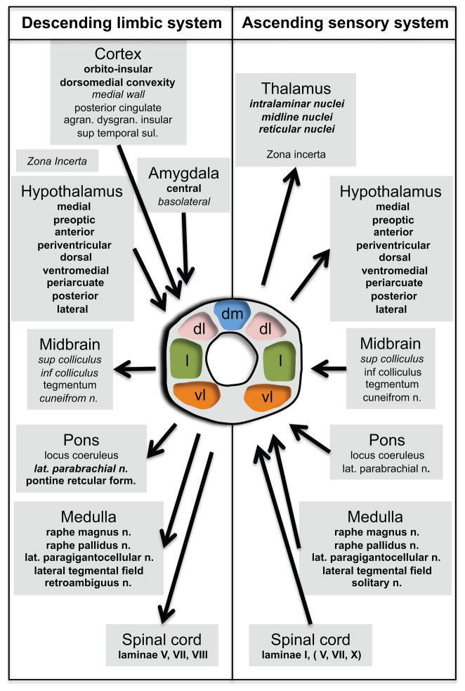
Figure 3. Anatomical Organization of the PAG.
Schematic overview of the organization of the PAG afferent and efferent connections. Represented on the left are the connections forming the descending limbic system and on the right are the connections forming the ascending sensory system. The two systems interact in the PAG. Structures indicated in bold are connected to either the dorsomedial, lateral or ventrolateral columns, or two of them or all of them. Structures indicated in italic are connected to the dorsolateral column. Structures indicated in bold and italic are connected to all four columns. The specific connections of the structures indicated in regular style have not been established. Adapted from (Paxinos and Mai, 2004), Figure 12.13 with permission.

