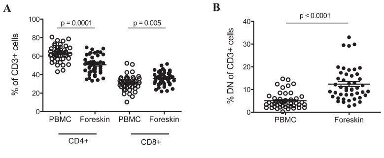
Figure 1. CD4+ and CD8+ T cell subsets within the foreskin and peripheral blood.
PBMC and foreskin cells from 46 men were stained with CD3-FITC, CD4-PE, and CD8-PerCP. Graphs show percentages of CD3+ cells within PBMC or foreskin cells that co-express (A) either CD4 or CD8, or (B) expressed neither CD4 nor CD8 (double negative, DN, T cells).

