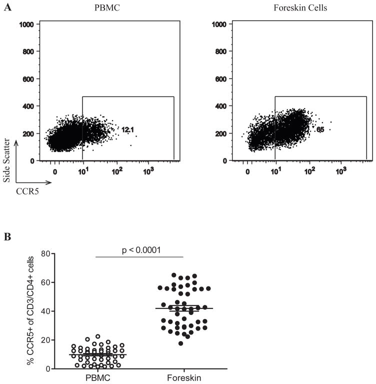
Figure 2. CCR5 expression on CD4+ T cells from the foreskin and peripheral blood.
PBMC and foreskin cells from 46 men were stained with CD3-APC, CD4-PE, and CCR5-FITC. Plots in (A) were created by gating on CD3+/CD4+ events. The gate defining CCR5+ events was created based on PBMC staining for this marker and applied to foreskin plots. (B) Proportions of CD3+/CD4+ cells in PBMC and foreskin cells co-expressing CCR5.

