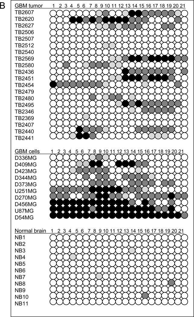
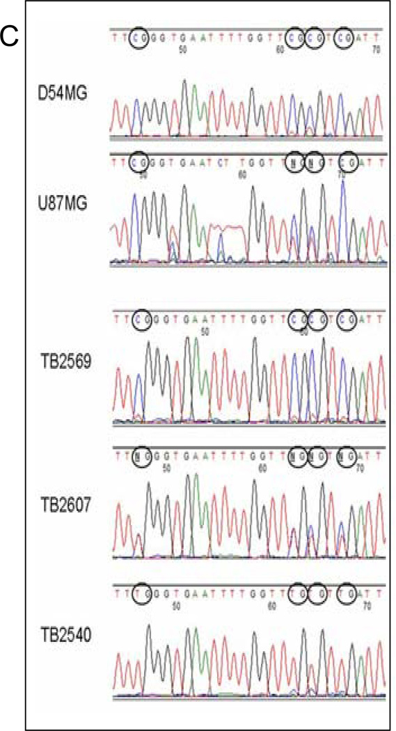
Figure 4.
Bisulfite treatment followed by methylation-sensitive quantitative PCR demonstrates extensive promoter methylation in AJAP1. A, schematic structure of the AJAP1 gene and location of candidate CpG islands within promoter region. *above red line = site of 5’ primers, *below red line = site of 3’ primers. B, summary of methylation-sensitive quantitative PCR analysis of 21 CpG sites in 20 primary glioblastoma tumors, 10 glioma cell lines and 11 normal brain samples. (Black circle >25% methylation; striped black circle = 10–25% methylation; gray circle <10% methylation; white circle = no methylation). C, representative sequencing chromatograms demonstrating CpG island methylation after bisulfite treatment in cell lines D54MG and U87MG, as well as tumors TB2569 and TB2607, and lack of methylation in TB2540.


