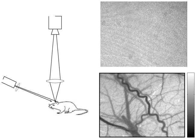
Figure 1.
(a) Basic laser speckle contrast imaging setup consisting of a laser diode and camera. A raw speckle image of the rat cortex, taken through a thinned skull, contains seemingly little information (b). However, when the speckle contrast is calculated using a sliding window (c), a tremendous amount of information is revealed about the motion of the scattering particles in the sample.

