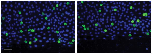
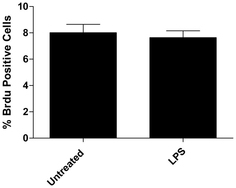
Figure 2.
A) Representative image of BrDU staining for cellular proliferation. The % FITC-positive cells were not significantly different in LPS treated as compared to untreated. B) Cellular proliferation not contributing to wound healing. Cells were counterstained with DAPI and images were taken of each wound using DAPI and FITC filters. Values are means of % FITC positive cells as compared to DAPI positive ± SD, with 4 wounds examined per treatment. Untreated 8.01% ± 2.2% vs LPS 7.64% ± 1.6%, NS.


