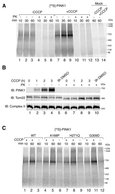
Figure 1.
In vitro import and BN-PAGE analysis of PINK1. (A) [35S]-PINK1 was incubated with isolated HeLa mitochondria with or without 1 μM CCCP for increasing times as indicated. Samples were treated with or without Proteinase K (PK) and solubilized in 1% digitonin containing buffer. Mock import samples lacking mitochondria were treated as above as indicated. (B) Mitochondria were isolated from HeLa cells that were either untreated or treated with CCCP or vehicle control (DMSO). Isolated mitochondria were treated with or without external protease (PK) and subjected to BN-PAGE and immunoblotting using antibodies against PINK1 (outer membrane), Tom22 (outer membrane) and Complex II (inner membrane). (C) Radiolabeled WT PINK1 and PINK1 patient mutants A168P, H271Q and G309D were imported into isolated HeLa mitochondria as in (A). Radiolabeled proteins were detected by phosphorimage analysis. See also Figure S1.

