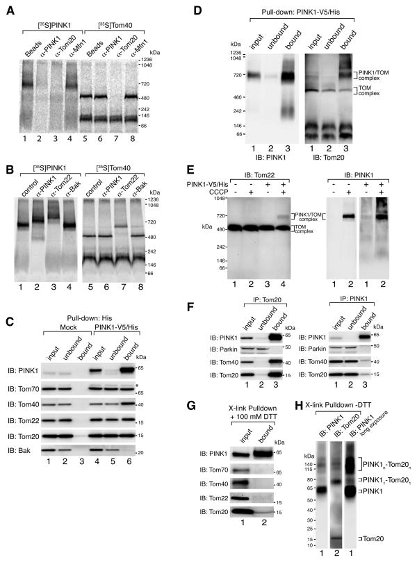
Figure 3.
PINK1 complex is associated with components of the TOM machinery. (A) Radiolabeled proteins were imported into isolated HeLa mitochondria in the presence ([35S]-PINK1) or absence ([35S]-Tom40) of 1 μM CCCP for 60 min. Samples were solubilized in 1% digitonin buffer and complexes were immunodepleted using indicated antibodies or beads alone as a control followed by BN-PAGE analysis. (B) Radiolabeled proteins were imported into isolated HeLa mitochondria in the presence ([35S]-PINK1) or absence ([35S]-Tom40) of 1 μM CCCP for 60 min. Samples were solubilized in 1% digitonin buffer followed by the addition of antibodies as indicated, and subjected to BN-PAGE. (C) Mock transfected and PINK1-V5/His stably transfected HeLa cells were treated with 20 μM CCCP for 3 h followed by mitochondrial isolation and immunocapture using α-His antibodies coupled to beads. Bound proteins were eluted with 6xHis peptides and various fractions as indicated were subjected to SDS-PAGE followed by immunoblotting using antibodies as indicated. *, non-specific band. (D) Samples treated as in (C) were analyzed using BN-PAGE and immunoblotting with α-PINK1 (left panel) and α-Tom20 (right panel) antibodies. (E) HeLa cells were either mock transfected or transfected with PINK1-V5/His and treated with or without 20 μM CCCP for 3 h before solubilization in 1% digitonin buffer followed by BN-PAGE and immunoblotting using α-Tom22 (left panel) or α-PINK1 (right panels) antibodies. (F) HEK293 cells were treated with 20 μM CCCP for 3h and then harvested and lysed in 1% digitonin containing buffer. Clarified lysates were used for immunopreciptation using α-Tom20 (left panel) or α-PINK1 (right panel) antibody coupled beads. Input, unbound and bound fractions were subjected to SDS-PAGE and immunoblotted using antibodies against PINK1, Parkin, Tom40 and Tom20. (G) Immunocaptured PINK1-V5/His complex as in (C) was incubated in 0.1 mM dithiobis[succinimidyl propionate] for 20 min on ice. After crosslinking, samples were incubated in 1% SDS containing buffer for 5 min at 95° and then subjected to immunoprecipitation using α-His beads. Crosslinker was cleaved with 100 mM DTT before SDS-PAGE and immunoblotting with antibodies as indicated. (H) Samples were treated as in (G) and subjected to SDS-PAGE in the absence of DTT followed by immunoblotting using α-PINK1 and α-Tom20 antibodies. Radiolabeled proteins were detected by phosphorimage analysis. See also Figure S2.

