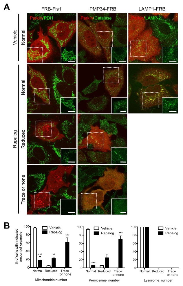
Figure 5.
Parkin recruited by ectopic PINK1 can induce autophagy of mitochondria and peroxisomes. (A) HeLa cells were transfected with PINK1Δ110-YFP-FKBP and cherry-Parkin together with one of the organelle specific FRBs (FRB-Fis1 (left column), PMP34-FRB (middle column) and LAMP1-FRB (right column)) for 24 h. After 48 h of treatment with (lower 3 rows) or without (top row) rapalog, cells were fixed, immunostained for organelle specific proteins (pyruvate dehydrogenase subunit E1α (PDH) for mitochondria (left column), catalase for peroxisomes (middle column) and LAMP-2 for lysosomes (right column)) and imaged with confocal microscopy. In rapalog treated cells, representative images of cells showing three different responses (normal, reduced and trace or none) of organelle mass are shown. White boxes on the right bottom corner show only the organelle markers from the dashed box to clearly show the change of organelle mass. White bar, 10 μm. (B) Cells having the indicated amount of each organelle in (A) were counted. To ensure the co-expression of the three constructs (PINK1-FKBP, Parkin and FRB) only the cells showing an abundant expression of mCherry-Parkin were counted. The graphs represent means ± SEM of counts in >100 cell per condition in three independent experiments and analyzed with 2 way ANOVA. ***; P<0.001, **; P<0.01. See also Figure S5.

