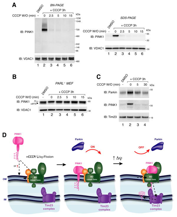
Figure 7.
PINK1 and Parkin after CCCP washout. (A) HeLa cells were treated with either DMSO or CCCP for 3 h before CCCP washout for increasing times as indicated. Cells were lysed in 1% digitonin buffer (BN-PAGE; left panel) or SDS sample buffer (SDS-PAGE; right panel) and immunoblotted using α-PINK1 and α-VDAC1 antibodies. (B) PARL−/− MEFs transfected with PINK1-V5/His were treated as in (A). Cells were lysed in SDS sample buffer and immunoblotted using α-PINK1 and α-VDAC1 antibodies. (C) HeLa cells were treated as in (A) and mitochondrial fractions were subjected to SDS-PAGE and immunoblotting using α-Parkin, α-PINK1 and α-Tim23 antibodies. W/O: washout, FL: full-length, ΔMTS: mitochondrial targeting sequence cleaved. (D) Model for PINK1 regulation. See discussion for description. See also Figure S7.

