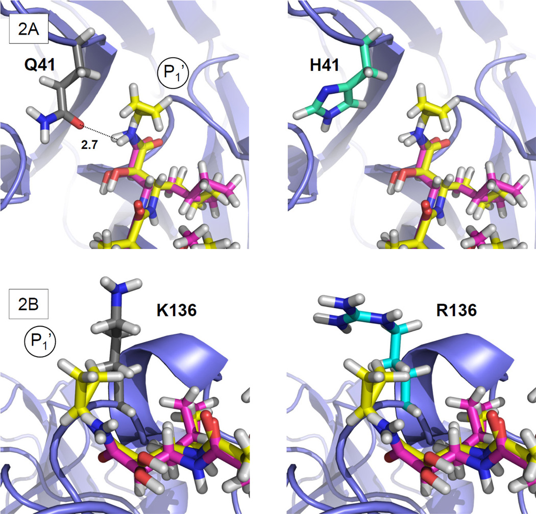
Figure 2. Rotamer analysis of Q41H and K136R.
Detail of PDB structure 2OC8 showing boceprevir (magenta) and the superposed TLL (yellow). The panels on the left show the side-chain conformation of the consensus residue, while those on the right show the predicted BSV conformation. (A) (left) The Q41 side-chain OH-group with predicted H-bond interaction (dotted line) with ketoamide backbone Cα, (right) this H-bond interaction is absent in H41. (B) Predicted R136 side-chain conformation and the TLL P1’ cyclopropyl showing closer van der Waals contacts in (right) R136 than in (left) the consensus K136.

