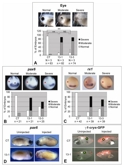
Fig. 1.
Knockdown of ADAM13 affects eye development in Xenopus. Eight-cell stage embryos were injected in one dorsal-animal blastomere with the indicated MO (1.5 ng), and cultured to desired stages. (A) Embryos were scored at stage ~35 for eye defects. One example of each phenotype is shown in the upper panels, and results of multiple experiments are graphed in the lower panels. **, p < 0.001 for comparison between control (CT) and 13-1 morphants, and p = 0.002 for comparison between CT and 13-3 morphants; NS, not significant (p = 0.91). (B and C) Embryos were cultured to stage ~12.5, and in situ hybridization was carried out for pax6 (B) or rx1 (C). The injected side is denoted with a red asterisk. e, eye field; l, lateral stripes. (D and E) Wild-type (D) or γ1-crys/GFP3 transgenic (E) embryos were injected with MO CT (upper panels) or 13-1 (lower panels). Embryos were cultured to stage 21–24 and processed for in situ hybridization for pax6 (D), or to stage ~45 (E). Representative embryos were photographed on both the uninjected (left panels) and injected (right panels) sides. Images taken in red (for co-injected red dextran) and green (for GFP) channels and bright field were merged in E. Note the lack of eye structures and GFP expression on the injected side of the 13-1 morphant. N = number of independent experiments; n = number of embryos scored (same below).

