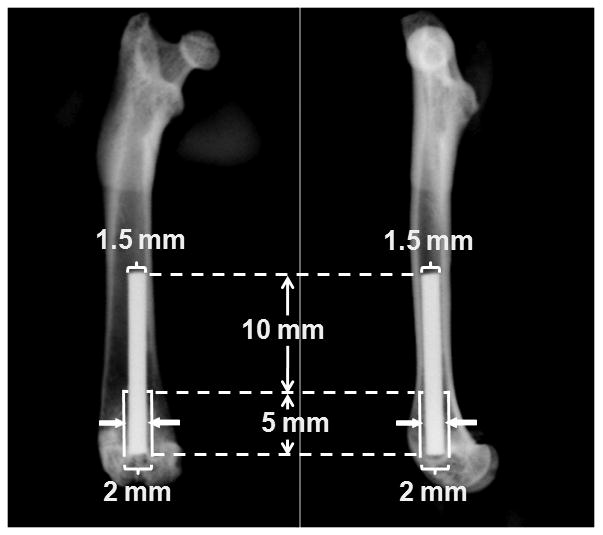
Figure 1.

Illustration of implant placement in the model. A 15 mm long by 1.5 mm diameter titanium implant was press fit in the distal end of the femur through the knee joint. A 0.25 mm wide gap (solid arrows) was created around the distal 5 mm of the implant, leaving good initial fixation at the proximal 10 mm of the implant.
