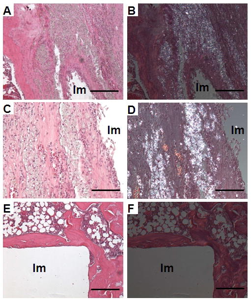
Figure 5.

Images show H&E staining under normal light (A, C, E) and under polarized light (B, D, F). Note the presence of a thick synovial-like membrane at the implant interface (A, C), associated with birefringent particles (B, D) in the particle-treated group. Samples in the control group showed a thick and even layer of peri-implant bone without a membrane at the bone-implant interface and a lack of birefringent particles (E, F). Scale bars in image A, B, E and F are 250 μm. Scale bars in image C and D are 50 μm. Im: Empty space after removing the implant.
