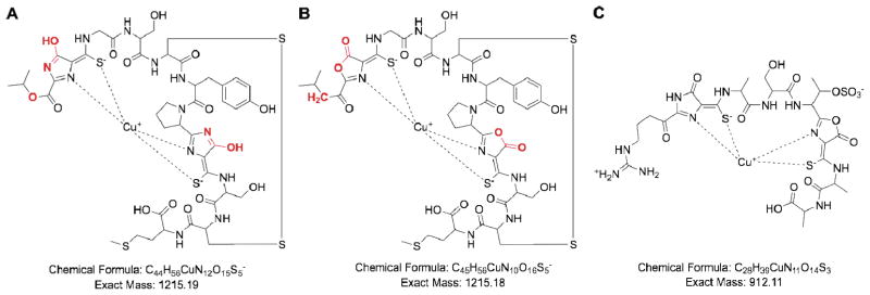
Figure 1.
Methanobactin structures. (a) Initial structure of M. trichosporium OB3b Cu-Mb determined by X-ray crystallography. (b) Revised solution structure of M. trichosporium OB3b Cu-Mb. Differences between the two structures are highlighted in red. (c) Proposed structure of Methylocystis strain SB2 Cu-Mb.

