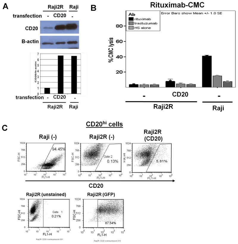
Figure 6. CD20 overexpression in RRCL indicated a potential defect in CD20 transport to cell surface.
20×106 Raji2R, Hela or Jurkat (data not shown) Tcells were transfected with 4μg of CD20-BCMGSneo vector by Amaxa Nucleofection. On day 2 or 4, cells were harvested for CD20 expression analysis by (A) Western blot. CD20 and β-actin control expression on Western blot were quantified by Image Jsoftware. (B) 51Crrelease assay was performed for rituximab-associated CMC. (C) Flow cytometry for percentage of cells with CD20 overexpression in mock-transfected and CD20-BCMGSneo vector-transfected Raji2R cells. Raji cells were used as positive control. Transfection efficiency was demonstrated post pmaxGFP transfection by GFP-expressing population.

