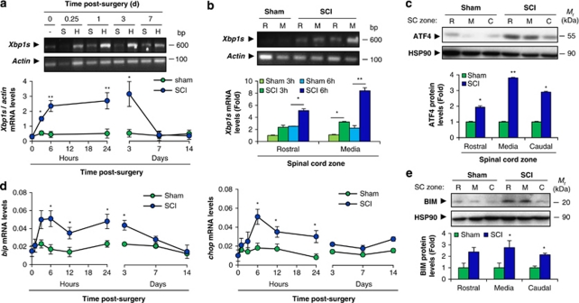
Figure 1.
UPR activation after spinal cord hemisection. (a) Wild-type mice were spinal cord-hemisected (H) or sham-operated (S) at the T12 vertebral level. 0.25 (6 h), 1, 3, 7, and 14 days after the surgical procedure, tissue from the operated region of the spinal cord was extracted and processed to measure spliced Xbp1 (Xbp1s) mRNA levels by RT-PCR. Actin mRNA was used for normalization. (b) The same procedure was used to study Xbp1 mRNA splicing at 3 and 6 h after SCI at the injured region (media) and at a distance from the injury site (rostral) (lower panel). Xbp1s levels were quantified and normalized using actin mRNA levels. (c) ATF4 protein levels were measured by western blot and semiquantified by normalizing with HSP90 protein levels. Blot and graph at 6 h after sham or hemisection from rostral (R), operated (M) and caudal (C) regions are shown. (d) bip and chop mRNA levels were quantified at the indicated times after damage by real-time PCR. (e) BIM protein levels at 6 h after SCI were measured by western blot. Protein levels were normalized with HSP90 levels and then normalized to the sham-operated condition. Mean±S.E.M. *P<0.05; **P<0.005; Student's t-test; n=3 animals per group for protein and mRNA analysis

