Figure 3.
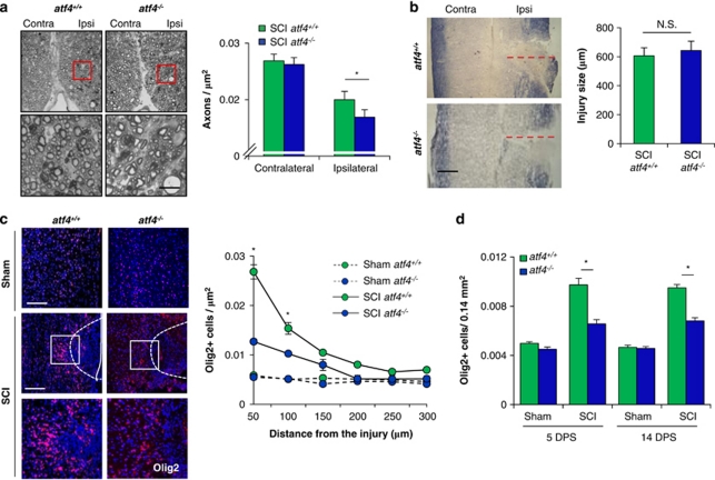
Altered cellular environment in ATF4-deficient mice after SCI. (a) atf4+/+ and atf4−/− mice were spinal cord-hemisected or sham-operated at the T12 vertebral level. At 14 days after surgery, spinal cord tissue was stained with toluidine blue to study axonal integrity (left panel). Intact axons from corticospinal tracts were quantified and expressed as axonal density values (right panel). (b) Spinal cord tissue from hemisected atf4+/+ and atf4−/− mice were longitudinally sectioned to analyze the injury size by eriochrome/cyanine myelin staining. Transversal injury length was measured from the edge of the spinal cord to the midline as shown by the red-dotted line. (c) atf4+/+ and atf4−/− mice were spinal cord-hemisected or sham-operated at the T12 vertebral level. At 5 days after surgery, spinal cord tissue processed for immunofluorescence for Olig2, to study oligodendrocytes and its progenitors (red); nuclei were counterstained using Hoechst (blue). Olig2-positive particles co-localizing with Hoechst were quantified every 50 μm starting at the injury site. (d) The same procedure as (c), but total Olig2/Hoechst-positive particles were quantified in a semicircular area of 300 μm radius surrounding the injury zone at 5 and 14 days post-surgery (DPS). Mean±S.E.M. *P<0.05; Student's t-test; n=3 animals per group. Scale bars, 20 μm in (a), 200 μm in (b), and 100 μm in (c). Bars indicate S.E.M.

