
Fig 1. The construction and characterization of pshA20-LMP1/2.
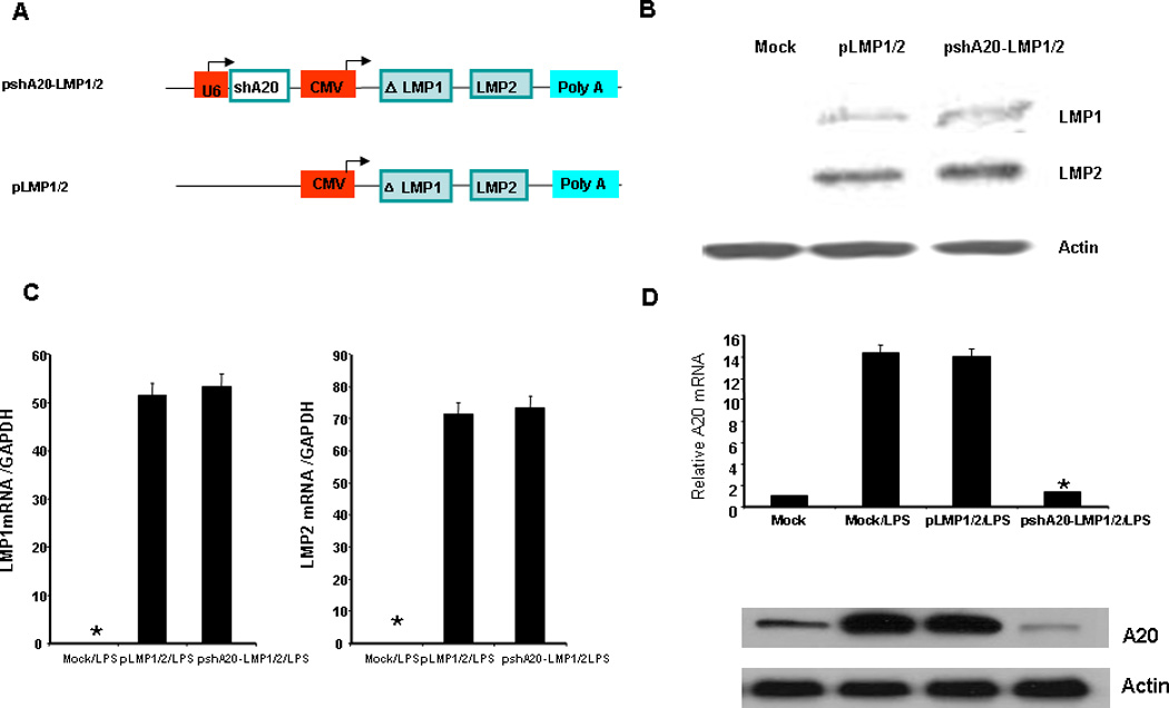
A. schematic representation of pshA20-LMP1/2 and control vector pLMP1/2. B. monocyte-derived DCs were nucleofected on day five with different vectors and matured with LPS (100 ng/ml) overnight. LMP1 and LMP2 expression in the DCs were analyzed 24 hours after nucleofection by western blot analysis. The antibody against EBV LMP1 or LMP2 was purchased from Santa Cruz Biotech (San Diego, CA). C. LMP1 and LMP2 mRNA levels were analyzed by qPCR 12hr after nucleofection. D. A20 mRNA level was analyzed by qPCR and data were normalized with mock DC A20 mRNA 24hr after nucleofection (upper). A20 protein expression was analyzed by western blot 24 hrs after nucleofection (lower). The data are representative of two independent experiments. *P < 0.01, pshA20-LMP1/2/LPS DCs vs. Mock/LPS DCs.