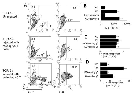
Fig. 6. Activated γδ T cells enhance the Th17 response in TCR-δ−/− mice.
(A) Three days before immunization with IRBP1-20/CFA TCR-δ−/− mice were left untreated (top panels) or were injected with 100,000 non-activated (resting) γδ T cells (center panels) or anti-CD3 antibody-activated γδ T cells (bottom panels) [33]. Fourteen days after immunization, 1.5 × 10⁶/well splenic T cells were stimulated with immunizing peptide and APCs in 24-well plates for 2 days and IFN-γ and IL-17+ T cells among the activated T cells were assessed by FACS analysis after intracellular staining for IFN-γ and IL-17. (B) Part of the culture supernatants was collected for IL-17 measurement. (C and D) LDA results showing the numbers of IFN-γ (Th1) or IL-17+ (Th17) T cells among 100,000 in vivo primed responder T cells. The results shown are representative of those in more than three experiments. In B–D, results for immunized WT B6 and TCR-δ−/− mice are shown for comparison.

